Dual PET imaging can connect tumor monitoring with heart-inflammation watchfulness
A dual PET approach could help researchers watch tumor activity and cardiac inflammation during cancer treatment, but PET signals need clinical correlation, validation and cardio-oncology boundaries before they guide care.
Ivy Stone ·
A dual PET-imaging idea is attractive because cancer treatment can create two urgent questions at once. Is the tumor becoming more active, and is the heart showing inflammatory stress that could change how safely therapy continues? The research item behind this article described a PET strategy meant to visualize both tumor progression and cardiac inflammatory responses during treatment. Its public value is not a promise of a new self-standing diagnosis. It is a clearer way to think about monitoring when oncology and heart safety overlap.
PET, or positron emission tomography, works by using a radioactive tracer that concentrates in tissues according to a chosen biological process. The familiar FDG tracer highlights glucose use, which is why many cancers and inflammatory tissues can appear active. Other tracers can be designed around receptors, metabolism or immune-cell behavior. In a dual-imaging approach, the point is not simply to make a brighter picture. It is to compare biological signals that may be moving in different organs while a treatment is being tested.
That distinction matters in cancer care. A therapy can shrink or slow a tumor and still affect the cardiovascular system; another apparent scan change may reflect inflammation, treatment effect, timing or technical factors rather than straightforward progression. Cardio-oncology exists for this borderland. It asks how to preserve cancer control while detecting heart injury early enough to adjust monitoring, supportive care or treatment plans under specialist supervision.
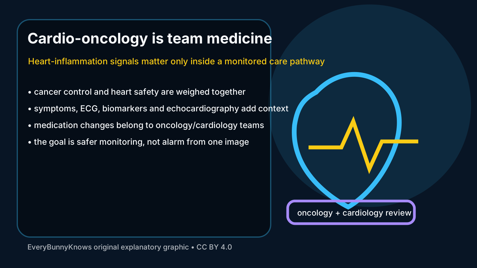
The strongest mechanism in a dual PET study is therefore integration. Tumor-facing images may show where malignant tissue is metabolically active or changing over time. Heart-facing images may show inflammatory activity that deserves correlation with symptoms, troponin or natriuretic peptide tests, ECG, echocardiography, MRI or clinical examination. One image can raise an important question; it should not be asked to answer every question alone.
The limits are especially important. Early PET work may involve small cohorts, preclinical models, selected patients or experimental tracers. A signal that is useful for research may not yet be reimbursed, standardized or available in ordinary hospitals. PET also has false positives and false negatives, exposes patients to a small amount of radiation and often needs CT or other imaging for anatomical context. Inflammation is a broad biological word, not a treatment decision by itself.
The safety boundary for readers is simple. This article does not advise anyone to start, stop, delay or choose cancer therapy, heart medicine or imaging. People receiving chemotherapy, immunotherapy, radiation or targeted treatment should discuss symptoms, scan results and cardiac risks with their oncology and cardiology teams. Chest pain, severe shortness of breath, fainting or sudden worsening symptoms need urgent medical care according to local services.
The hopeful part is practical rather than dramatic. Better imaging can give teams an earlier, more structured conversation: whether a tumor signal is changing, whether the heart needs closer attention, and whether a trial therapy is helping one problem while creating another. If larger studies validate the approach, dual PET could become one more careful tool in a monitored pathway where cancer control and heart protection are treated as connected goals.