Uneven cerebellum aging offers a clue to cognitive resilience in later life
A Nature Neuroscience study of brain imaging from about 47,000 adults found that cerebellar regions do not age uniformly and that larger cerebellar volume was linked with better cognitive scores. It is a clue about resilience, not a recipe for preventing decline.
Klára Novák ·
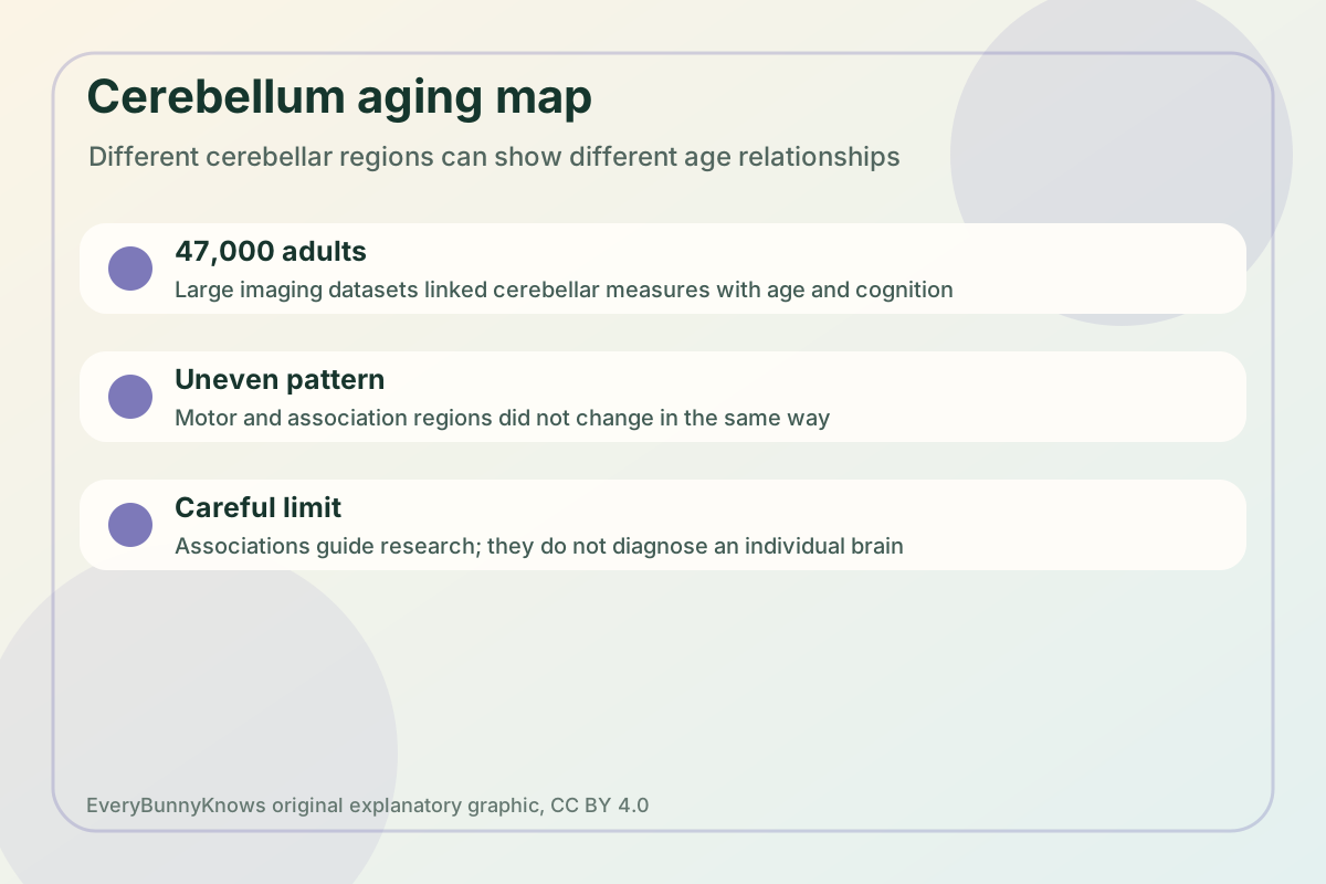
Aging research has often treated the cerebellum as a supporting actor: the small, tightly folded structure at the back of the brain that helps coordinate movement and balance. A 2026 paper in Nature Neuroscience makes the picture richer. Using brain-imaging data spanning about 47,000 adults, the authors reported that the cerebellum does not age as one uniform block. Some regions showed steeper age-related differences than others, and cerebellar measures were linked with cognitive performance in later life.
That matters because the cerebellum contains most of the brain’s neurons and connects with motor, sensory and association networks. The study used volumetry and the T1-weighted/T2-weighted MRI ratio, then checked parts of the pattern with quantitative MRI in an independent sample. In plain terms, the researchers were asking whether regional size and tissue-sensitive imaging signals form a map of cerebellar aging, and whether that map helps explain why some older adults keep stronger thinking skills than others.
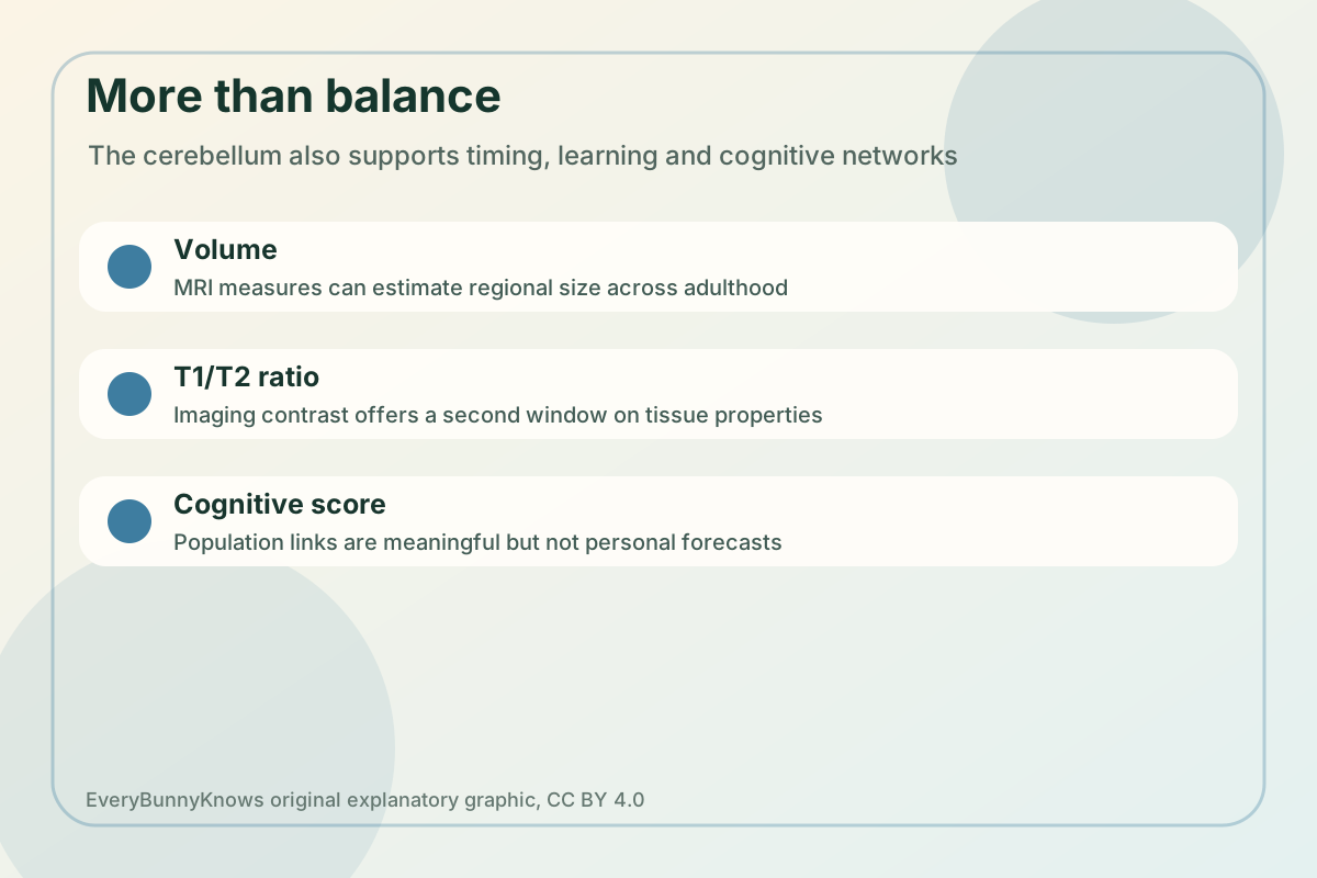
The mechanism is not that one brain area single-handedly preserves memory. The cerebellum helps tune timing, prediction and error correction across circuits. Regions connected more strongly with association networks may therefore contribute to attention, language or executive tasks, while motor-related lobules may follow a different aging curve. If one region loses volume faster, or if tissue properties shift in a pattern that tracks age, the effect may be visible in group-level cognitive scores even when any one person’s daily life is shaped by many other factors.
The study does not turn the cerebellum into a single key to successful aging. MRI correlations cannot prove that cerebellar differences cause better cognition, and a scan is not a personal forecast. The datasets are large, but imaging studies still depend on who was recruited, how scanners were harmonized, which cognitive tests were used and how health, education, vascular disease, hearing, mood and sleep were measured. Cognitive resilience is a whole-body and social story as well as a brain story.
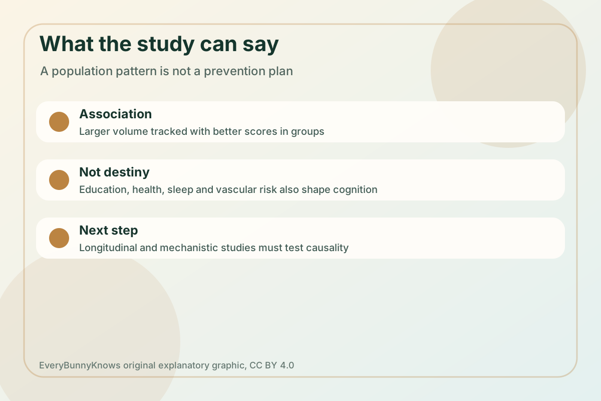
The careful value is in the map. A more detailed cerebellar atlas can help researchers ask sharper questions about which circuits are vulnerable, which remain stable and how exercise, cardiovascular health or neurological disease might interact with cerebellar structure. It may also nudge public understanding away from a narrow “balance center” view of the cerebellum.
For readers, the safest takeaway is curiosity rather than self-diagnosis. Brain aging is influenced by vascular health, education, sensory loss, social connection, medications, disease and chance, and the paper does not test an intervention. What it does offer is a better anatomical vocabulary, one that treats brain resilience as a network property rather than a vague trait. If future longitudinal studies can follow the same people over time, researchers may learn whether cerebellar changes precede cognitive shifts, accompany them or reflect compensation by networks that still work well. The hopeful part is modest but real and it gives later studies sharper clinical research targets: when aging is measured region by region, scientists get more chances to separate normal change from risk, compensation and resilience.